
Most full-size DEXA scanners support a maximum patient weight of about 227 kilograms (500 pounds), according to the manufacturer pre-installation specifications for GE Lunar's iDXA. Older or compact models often cap out lower, closer to typical weight ranges seen in clinical reviews, while some specialty systems claim more. Before you book, call the clinic and confirm the exact model, its rated weight limit, and its table width.
TL;DR:
- Weight limits for full-size DEXA scanners usually max out at around 227 kilograms (500 pounds), but older models often support closer to 159 kilograms (350 pounds).
- Scan window width, typically between 58 and 65 centimeters, can prevent larger patients from fitting, even if their weight is below the rated limit.
- Exceeding a scanner’s size or weight capacity can reduce measurement accuracy, especially for bone density, and often requires alternative protocols like half-body or peripheral scans.
- Confirm the specific model’s weight limit and table dimensions before booking to ensure it can accommodate your size, and consider measuring your body girth for a better fit assessment.
- When in doubt, ask clinics if they have higher-capacity machines, use positioning aids, or recommend alternative scans to get reliable results.
Table of Contents
- Manufacturer limits and typical ranges: what the spec sheets show
- Table width and scan-window clearance matter as much as weight
- What happens to accuracy when you exceed a scanner's limits
- Scan preparation, artifacts, and contraindications that can skew results
- What to confirm with the clinic before you schedule
- If you exceed a machine's limits: your options and their tradeoffs
- How Dexascans helps you find a machine that fits
- A short note on getting this right the first time
- Find a DEXA clinic that fits your size and budget
- Sources
- FAQ
Manufacturer limits and typical ranges: what the spec sheets show
The number you get quoted for a "DEXA scan weight limit" depends entirely on which machine sits behind the question. GE Healthcare's pre-installation documentation for the Lunar iDXA lists a maximum patient weight of 227 kilograms (500 pounds), a figure that has become something of an industry benchmark for modern fan-beam densitometers. Hologic's Horizon and Discovery series are typically listed in the same range, around 226 to 227 kilograms. That consistency is not a coincidence: both manufacturers built their current-generation tables around similar structural load ratings and similar clinical demand for scanning larger patients.
The picture looks different once you step back a model generation. GE's older Prodigy system, still common in many clinics and research settings, tops out closer to 159 kilograms (350 pounds). Hologic's older QDR series shares a similar profile. A clinical review of body composition measurement in overweight individuals found that weight limits across a range of densitometers, old and new, cluster between roughly 114 and 159 kilograms (250 to 350 pounds), with table widths in the range of 58 to 65 centimeters. Newer fan-beam systems tend to sit above that range, which is part of why a clinic's answer to "what's your weight limit" can vary so widely depending on the age of its equipment.

It helps to understand why the numbers vary at all. A manufacturer's stated weight limit is generally the safe working load for the scanning table and its motorized components, not a reflection of image quality at that weight. A table might mechanically support 227 kilograms without any problem, but the scan window, meaning the width the X-ray detector arm can pass over, was engineered around an average adult frame. That distinction matters because a patient can be well under the rated weight limit and still not fit within the usable scan area, a problem covered in the next section.
Some clinical literature references even higher figures for select systems, particularly bariatric-specific or research-grade units, though these are less common in routine outpatient settings. When a number sounds unusually high, ask the clinic whether it reflects the table's mechanical rating or the manufacturer's recommended scanned mass for accurate results. Those two figures are not always the same, and a clinic quoting the higher of the two without qualification is not giving you the full picture.
If you want to check a specific machine's specifications before calling, manufacturer sites and technical spec sheets are the most reliable source. Dexascans' own manufacturer comparison page breaks down current models from Hologic, GE, Osteosys, and Norland side by side, and its GE-specific brand page covers the Prodigy and iDXA lines in more detail, including the weight and table dimensions each one supports. Clinics themselves should also be able to tell you the model on request. If a receptionist cannot name the manufacturer or model, that is worth noting before you commit to an appointment.
Table width and scan-window clearance matter as much as weight
Weight capacity gets most of the attention, but it is often not the limiting factor for larger patients. The scan window, the physical width the detector arm travels across, is fixed by the machine's design and cannot flex the way a person's body can. The same clinical review that reported weight limits between 114 and 159 kilograms also found table widths clustering around 58 to 65 centimeters (roughly 23 to 26 inches) across several common densitometer models. A patient with a chest or hip girth that exceeds that width will have soft tissue extend past the edge of the scan area regardless of their total body weight.

This is why two people of the same weight can have very different experiences on the same machine. Someone who carries weight with a narrower frame may scan cleanly within the window, while someone with a wider chest or hip girth at a lower body weight may not. Vertical clearance under the scanning arm adds a second constraint: taller patients or those with a deeper chest wall can occasionally bump against the arm's travel path, though this is less commonly reported than lateral overhang issues.
Newer fan-beam systems generally offer more forgiving scan windows than older pencil-beam machines, which is part of why clinics with recent equipment can often accommodate patients that older machines cannot, independent of the weight rating itself.
Before you call a clinic, it is worth taking rough measurements of your own body:
- Chest girth, measured at the widest point around the ribcage.
- Hip and thigh girth, measured at the widest point while standing normally.
- Height, measured without shoes.
Compare these against the table width and length the clinic reports for its machine. If your chest or hip girth is close to or exceeds the stated scan-window width, ask directly whether the technician has experience positioning larger patients or whether a half-body protocol might be needed, a technique covered next.
What happens to accuracy when you exceed a scanner's limits
Exceeding a machine's practical limits, whether by weight or by girth, does not always mean the scan simply fails. More often, it changes what the scan measures and how reliable that measurement is. A technical review of DXA artifacts notes that excess soft tissue increases image noise, which reduces the precision of lumbar spine bone density readings and can bias trabecular bone score calculations. In practical terms, the scan may still produce a number, but that number carries more uncertainty than it would for a patient who fits comfortably within the scan window.
When a patient's body is too wide for the standard scan window, many clinics fall back on a half-body scan. The technician scans one side of the body and either mirrors the result or uses a validated extrapolation method to estimate total-body composition. This approach is accepted in clinical practice, but it comes with known error margins, since it assumes a degree of symmetry between the left and right sides of the body that is not always accurate. A half-body result should be treated as a reasonable estimate rather than a precise total-body figure, and it is worth asking the clinic to note on your report that a half-body protocol was used, so any future comparison accounts for that difference.
For bone density specifically, forearm or peripheral DXA offers another workaround when a central scan (spine or hip) is not feasible due to size, positioning, or hardware artifacts. Peripheral scanning uses a smaller, separate device to measure bone density at the forearm or heel. It is faster and imposes far fewer size constraints than a full-body table, but it measures a different skeletal site than the hip or spine, so the result is not interchangeable with a central DXA reading for diagnosing osteoporosis. The updated clinical practice guideline for DXA notes that weight limits vary by scanner type and recommends measuring weight and height at the time of each scan, using alternative sites when a central scan is compromised.
None of these workarounds are failures. They are established clinical accommodations, and a technician who suggests one is generally trying to get you the most reliable measurement available given the equipment on hand.
Scan preparation, artifacts, and contraindications that can skew results
A DEXA scan is sensitive to more than just weight and body size. Several everyday factors can distort the reading if you do not prepare correctly, and a few medical situations rule the scan out entirely for a period of time.
Standard patient preparation guidance from Massachusetts General Hospital's DXA instructions recommends the following before a scan:
- Remove metal objects, including jewelry, belts, and any clothing with metal fasteners or underwire.
- Avoid calcium supplements for about 24 hours before the appointment.
- Tell the technician about any internal metal hardware, such as joint replacements, spinal rods, or surgical clips.
- Confirm you are not pregnant, since pregnancy is a contraindication for DEXA scanning.
- Mention any recent imaging studies that involved contrast dye.
That last point deserves its own explanation. Oral or intravenous contrast agents used in CT scans or other imaging can temporarily elevate measured bone density. A review of DXA artifacts describes case reports where iodine-based contrast inflated BMD readings, with the artifact resolving in repeat scans taken roughly 7 to 13 days later. The same source and the Massachusetts General Hospital guidance both point to a waiting period in the 7 to 14 day range after a contrast study, depending on the type of contrast used, before a DEXA result can be considered reliable.
Statistic Callout: Recent contrast imaging can temporarily distort bone density readings, and waiting roughly 7 to 14 days after a contrast study is commonly recommended before scheduling a DEXA scan. That single scheduling detail can be the difference between an accurate baseline and a misleading one.
Internal hardware, oral contrast still in the digestive tract, ports, and vascular calcifications can all change measured BMD at the specific vertebrae or region they overlap. Clinical guidance from the International Society for Clinical Densitometry, as referenced in artifact literature, allows technicians to exclude affected vertebrae from the analysis and interpret the remaining evaluable sites, or switch to a forearm scan when the lumbar spine and hip are both compromised. If you have any hardware or a recent procedure involving contrast, mention it when you book, not just when you arrive.
What to confirm with the clinic before you schedule
A short phone call before booking can save you a wasted appointment. Here is what to ask, in order:
- What is the scanner make and model? This tells you which spec sheet applies to your case.
- What is the maximum patient weight, in pounds or kilograms? Ask for the number specific to that model, not a general industry figure.
- What is the table width? This matters more than weight for many larger patients, as covered above.
- Do you offer a half-body protocol for patients who don't fit the standard scan window? Not every clinic does, and it is worth knowing in advance.
- Can you note the machine model on my scan report? This matters for anyone tracking bone density or body composition over time, since comparing results across different manufacturers' equipment introduces its own inconsistencies.
It is also reasonable to ask how often the technician works with larger patients and whether they use positioning aids, such as straps or foam wedges, to reduce lateral overhang during the scan. Experienced technicians often have practical strategies for getting a valid scan even when a patient is near the edge of the machine's limits.
Pro Tip: If a clinic can't quote your model's exact weight limit or table width over the phone, ask them to send a photo of the machine or a copy of its spec sheet before you book.
Since DEXA scans booked through independent clinics typically do not require a physician referral, it is worth confirming that upfront as well, along with how the clinic handles payment and whether pricing is disclosed before your visit.
If you exceed a machine's limits: your options and their tradeoffs
If a nearby clinic's equipment genuinely cannot accommodate your size, you have a few reasonable paths forward. The most direct is asking for a referral to a hospital radiology department or a research center, both of which sometimes operate higher-capacity or bariatric-specific densitometers not commonly found in standalone imaging clinics. These facilities are more likely to have newer fan-beam systems built with a wider scan window in mind.
Portable densitometers are another option, though availability varies significantly by region and the units themselves differ in their capacity and scan quality compared to full-size central DXA tables. Dexascans' guide to portable DEXA and bone density machines covers which models exist and how they compare to stationary systems, useful context if a portable option comes up in your search.
When central DXA truly is not workable, alternative methods exist for tracking body composition or bone health, each with real limitations. Bioelectrical impedance analysis (BIA) estimates body fat using electrical resistance and is widely available but generally less precise than DXA. CT-based body composition analysis can measure fat and lean tissue with detail, though it involves radiation exposure well above a standard DEXA scan and is rarely used outside research or specific clinical indications. Simple anthropometry, tape-measure girths and skinfold calipers, is the least precise of the group but requires no special equipment. None of these fully replace DXA's role as the standard for bone mineral density, and most clinicians treat it as the reference point even when a patient needs a workaround to get scanned.
How Dexascans helps you find a machine that fits
Finding a clinic that can actually accommodate your size and needs is largely a matter of information, not luck. Some platforms list verified DEXA locations across many states, often displaying cash pricing upfront to help users compare options before contacting clinics. That verification process is what makes it practical to narrow down clinics before you ever pick up the phone.
A few ways to use the platform when weight or size is a concern:
- Look at the portable DEXA machine guide if a fixed clinic table is not a good fit and a mobile option might work better.
- Compare listed scan types, since some locations offer combined body composition and bone density appointments while others specialize in one or the other.
The goal is not to guess which local clinic has the right equipment. It is to check verified listings first, then confirm the specific model and table dimensions directly with the clinic using the checklist above. That combination, transparent information plus a short confirmation call, is the most reliable way to avoid an appointment that does not work for your body.
A short note on getting this right the first time
If there is one thing worth taking away from all of this, it is that the phone call before booking matters more than the general statistics. Weight limits and table widths vary enough between models that a number you read online, including the ones in this article, may not match the specific machine at your nearest clinic. Measuring your own chest and hip girth takes a few minutes and gives you something concrete to compare against whatever the clinic tells you.
If a machine turns out not to be a good fit, that is not a dead end. Half-body protocols, forearm scans, and referrals to higher-capacity centers all exist precisely because this situation comes up regularly. Asking the clinic to document the model on your report also pays off later, since sticking with the same type of equipment makes any future comparison more meaningful.
— Erik
Find a DEXA clinic that fits your size and budget
Once you know what to ask, the fastest path forward is comparing clinics that have already been verified rather than calling around one by one. Users can find verified DEXA scan locations with transparent cash pricing shown before booking, allowing cost comparison without needing to call clinics individually.
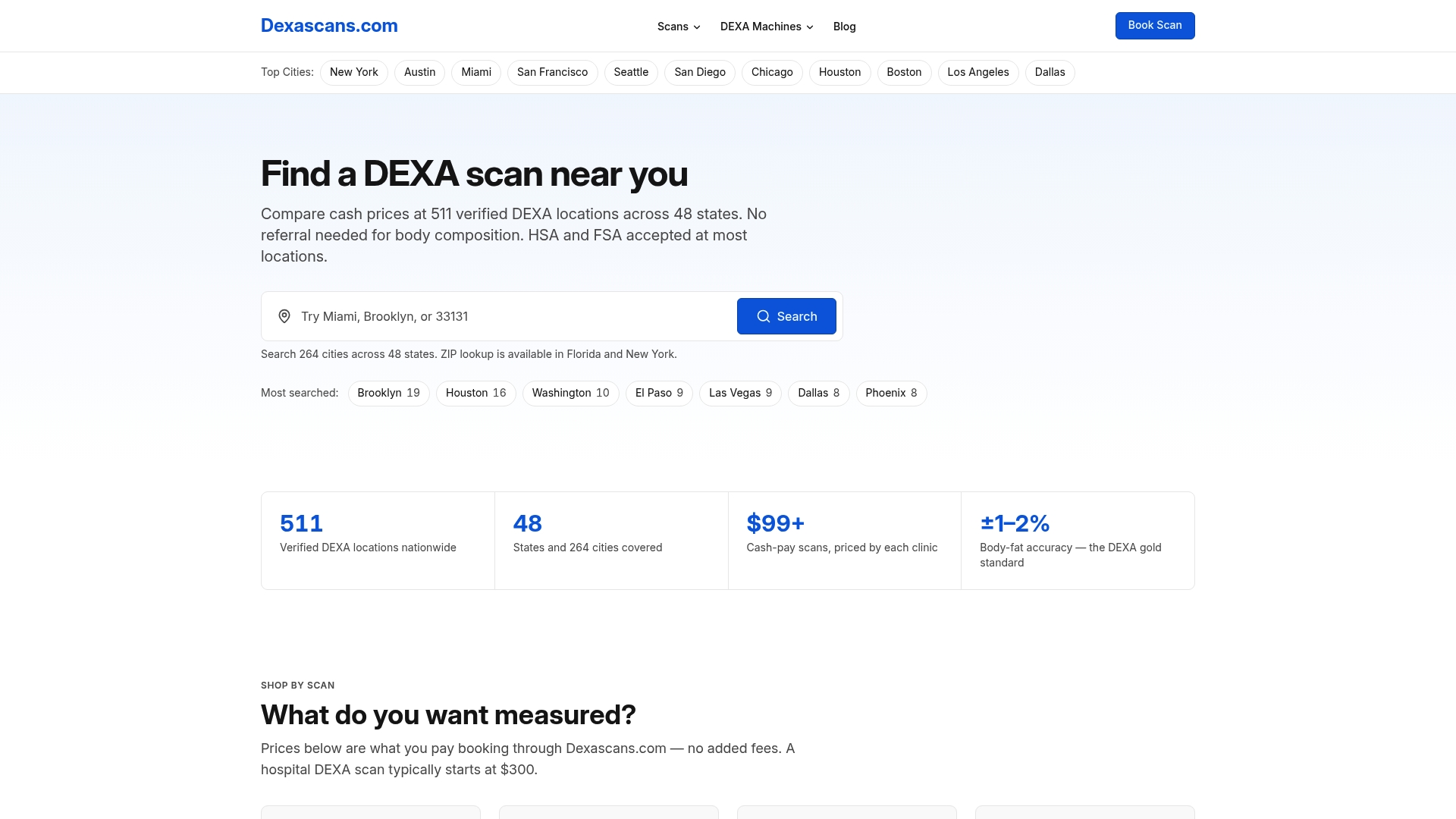
Search the locations page by city or ZIP code to compare nearby clinics, their scan offerings, and their pricing side by side. If you want to check machine specifications before you call, the manufacturer comparison page breaks down current models by brand, giving you a starting point for the exact questions covered above.
This article is general information, not a substitute for advice from a qualified doctor. Consult a qualified healthcare professional about your own circumstances before acting on anything here.
Sources
- Pre-installation Manual and Checklist for Prodigy and Lunar iDXA
- Measuring body composition in overweight individuals by dual energy x-ray absorptiometry (DXA)
- Patient instructions for DEXA (Massachusetts General Hospital / TCRC resource)
FAQ
What can throw off a DEXA scan?
Metal jewelry or clothing fasteners, recent calcium supplement use, and internal hardware like joint replacements can all interfere with a scan. Recent contrast dye from another imaging study can also temporarily raise measured bone density, which is why waiting about 7 to 14 days after a contrast study is commonly advised.
Can I wear deodorant during a bone density scan?
Deodorant itself is not typically listed as a concern for bone density scans, but the standard preparation guidance focuses on removing metal objects and clothing with metal fasteners before the appointment. If you are unsure about a specific product, ask the clinic directly when you book.
Who cannot have a DEXA scan?
Pregnant patients should not have a DEXA scan, since the standard patient guidance lists pregnancy as a contraindication. Patients who recently had a contrast imaging study or who exceed a specific machine's weight or width capacity may also need to wait or use an alternative scanning approach.
What are the downsides of a DEXA scan?
Patients whose body size exceeds a scanner's table width can experience image noise and reduced precision in the results, particularly for lumbar spine measurements, according to technical reviews of DXA artifacts. Internal hardware, recent contrast dye, and certain positioning limitations can also affect accuracy if not addressed before the scan.
How do I know if a clinic's machine can accommodate my weight?
Ask the clinic for the exact scanner make and model, then compare its rated weight limit and table width against your own measurements before booking. You can also check verified clinic listings and manufacturer specs on the Dexascans locations page before making a call.